x-pur crystal bottle
In-Office Desensitizer

Ingenious, natural and fast-acting desensitizer

Professionals

Description

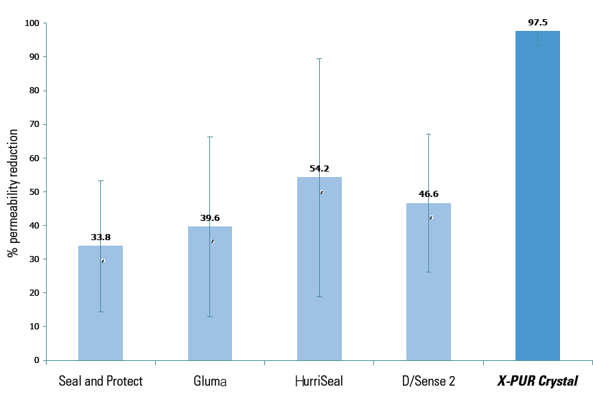
X-PUR Crystal is a water-based potassium oxalate in-office desensitizer made of natural acids and salts that quickly form insoluble calcium crystals. It provides effective patient relief for the general root, or exposed dentin sensitivity, in only a few seconds. This innovative blend rapidly blocks the fluid flow to help provide relief from sensitivity. It is uniquely different than other desensitizers on the market because it does not have to be refrigerated. For extra patient comfort, it can be warmed to body temperature or slightly above. X-PUR Crystal is efficient, cost-effective and safe.

Mechanism of action

Enamel, dentin and cementum are composed of high concentrations of alkaline calcium hydroxyapatite (HAp). X-PUR Crystal reacts with HAp to rapidly form millions of acid resistant chelate precipitates within the dentinal tubules at depths of 7-12 μm and on the surfaces of the vital dentin substrate, this action creates a long-lasting seal, preventing patient hypersensitivity and blocking bacteria penetration to the vital pulp.

Benefits

  • Biologically compatible with soft-tissue, works well after periodontal surgery
  • Can be used with any adhesive systems
  • Cost-effective: $0.31 per drop, 1-2 drop(s) per site. 10ml bottle = 320 drops
  • Easy application: no drying, isolation or curing necessary
  • Glutaraldehyde and HEMA free: non-irritating and no harm to soft-tissues
  • Long-lasting, quick and easy application (20 sec. saturate + 20 sec. rest)
  • May enhance tissue re-attachment
  • Prevents the migration of bacteria towards the vital pulp
  • Slows the demineralization process and promotes remineralization

Indications

  • Crown preparation: before and after
  • Exposed cementum, dentin and root surfaces
  • Heavy calculus along the cervical neck that extends along the root surface
  • Pain from removal of an orthodontic bracket
  • Periodontal surgery
  • Prophylaxis: before and after
  • Re-wetting agent
  • Temporization
  • Thermal sensitivity
  • Under restorations: amalgams, composites, crowns and veneers, inlays and onlays
  • Whitening: before and after

Ingredients

Proprietary blend of natural acids and salts

Clinical uses

  • Periodontitis
  • Tooth sensitivity
  • Whitening

Directions for use

Do not etch X-PUR Crystal’s low pH formulation. It dissolves immediately, does not harm soft-tissues, and effectively removes the smear layer.

Adults

  1. Clean and rinse the area of all foreign debris of biofilm or other particles without a gauze. *It is best to not dry as the product is hydrophilic. Note: using a gauze to rub the area may desiccate the dentinal tubule.
  2. Place 1 to 2 drop(s) for every 1-2 sites into a clean well of your choice or a dappen dish. You may need more drops depending on the size and number of teeth.
  3. Using a sterile micro brush, microsponge or small cotton pellet, soak the area and gently rub it onto the affected surface(s) for 20 seconds. Note: ensure to continually dip the brush into the liquid and rub into the area (back/forth) to thoroughly saturate the exposed area.
  4. Allow X-PUR Crystal to naturally air dry only for 20 seconds. Note: Keep the buccal and labial mucosa off the teeth after applying to aid in air drying.
  5. If you do not experience the desired relief, you may perform a second or third application without any tissue damage.

Directions for Use Under a Restoration or Crown

  1. It is suggested not to etch under a restoration because of it’s low pH formulation. X-PUR Crystal is considered a self-etchant which dissolves immediately and already removes the smear layer.
  2. If you want to etch: Etch, apply, prime, and bond.
  3. Place when all of the restorative drilling is completed.
  4. If you wish to place an adhesive restoration, place an acid etchant for only 5 seconds to remove the debris, then rinse and gently evacuate with light air dispersion of 1 second and at a distance of 5-cms.
  5. Apply X-PUR Crystal onto all of the preparation surface as well as cavity walls and even onto the enamel around the cavity prep.
  6. Apply the X-PUR Crystal with a microsponge brush and be sure to rub the agent all over the cavity prep surfaces: This is to ensure the agent contact is complete, to permit chelation of many millions of small nano-Calcium oxalate acid resistant crystals to seal the dentinal tubules, as well as, to create a surface layer that does not diminish adhesive bond strengths.
  7. After 20 seconds of rubbing X-PUR Crystal, do not rinse, but evacuate the area with gentle suction and 1 second air puffs from 5 cm distance.
scanning electron microscope image

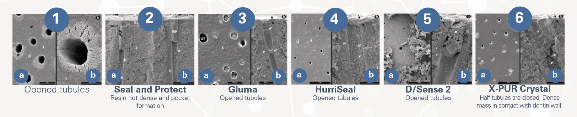
* Scanning Electron Microscope (SEM) image of the very dense peritubular dentin (calcium hydroxyapatite) and the very small crystals along the inner wall which are insoluble calcium crystals that assist to relieve patient discomfort.

Clinical tips

Any appearance of a frosty-white surface indicates the blockage of the dentin tubules. May be applied to a damp surface and dabbed around crown margins and onto the exposed tooth roots that are sensitive to cold or air stimuli. No light curing is necessary.

Reduces cariogenic bacteria load of highly infected patients

Reasons X-PUR Crystal Glutaraldehyde + HEMA
Desensitizing effect* 97.5 46.6
Application time 30-40 sec 60 sec
Interference with bonding systems (Bonding takes place in the intertubular dentin) No
Act only in the peritubular dentin
Possible
Phosphoric acid etching formulas may affect resorations
Interference with self-etch systems No Not recommended
Irritation potential No Yes
Rubber dam isolation No Yes
Rinsing required No Yes

% permeability reduction of the dentin desensitizers tested.

Effect of desensitizing agents on dentin permeability and dentin tubule occlusion.

Justine L. Kolker / Marcos A. Vargas / Steven R. Armstrong / Deborah V. Dawson, J Adhes Dent. 2002 Fall;4(3):211-21.
  1. Scanning electron micrographs (SEM) of polished dentin. No smear layer or smear plugs are observed on the dentin. a. A lateral canal is observed within the intertubular dentin and is also devoid of smear-layer and plug (black arrow). P: peritubular dentin; I: intertubular dentin. Micron bar figure a = 5μm. Micron bar figure b = 2μm.
  2. SEM of Seal and Protect, fractured specimen. A resin layer (RL) covers the surface and infiltrates the tubules with a resin plug (RP). Globular structures are present below the RP within the dentinal tubules (white arrows). The RP occluding the tubule shows a pocket formation (black arrows). P: peritubular dentin; I: intertubular dentin. Micron bar = 5μm.
  3. SEMs of Gluma. a. top surface. A thin coat (C) is seen over the dentin. Some of the tubules appear to be open (OT) and some appear closed (CT). Micron bar = 2μm. b. fractured specimen. A thin coat covers the dentin surface (black arrows) it extends within the tubules covering the walls (black open arrows) and forming bridges between them (white arrows). P: peritubular dentin; I: intertubular dentin. Micron bar = 1μm.
  4. SEMs of HurriSeal. a. top surface. Most of the dentinal tubules appear to be open. Micron bar = 10μm. b. fractured specimen. A coat covers the dentin surface (black arrows). Globular structures are present within the dentinal tubules (white arrows). P: peritubular dentin; I: intertubular dentin. Micron bar = 1μm.
  5. SEMs of D/Sense 2. a. top surface. A crystal precipitate (white arrow) is seen covering part of the dentin. Tubules that can be seen appear open. Micron bar = 10μm. b. fractured specimen. A thin coat covers the dentin surface (black arrows). Within the tubule crystals (white arrow) form a fairly dense mass, yet do not appear to be attached to the tubules (black open arrows). An open lateral canal free of crystals is marked with a white *. P: peritubular dentin; I: intertubular dentin. Micron bar = 1μm.
  6. SEMs of X-PUR Crystal. a. top surface. About half of the dentinal tubules appear to be closed. A few crystals are apparent on the dentin surface (white arrow). Micron bar = 10μm. b. fractured specimen. A thin coat covers the dentin surface (black arrows) and invaginates (white *) into the dentinal tubule. Below the invagination crystals have formed a dense mass (white arrows) and have intimate contact with the dentin wall (open black arrows). Micron bar = 1μm.

Featured publications

The effect of oxalate-based desensitizers on the microleakage and shear bond strength of resin modified glass ionomer

The effect of oxalate-based desensitizers on the microleakage and shear bond strength of resin modified glass ionomer

X-PUR CRYSTAL: THE SCIENCE BEHIND

X-PUR CRYSTAL: THE SCIENCE BEHIND

Effect of desensitizing agents on dentin permeability and dentin tubule occlusion.

Effect of desensitizing agents on dentin permeability and dentin tubule occlusion.